FORMATION OF 2 DIMENSIONAL PROTEIN CRYSTALS FOLLOWED BY ATOMIC FORCE MICROSCOPY
Ilya Reviakine, Wilma Bergsma-Schutter, Alain Brisson
Electron Microscopy group Biophysical Chemistry,
University of Groningen, Nijenborgh, 4, 9742 AG Groningen, the Netherlands
Keywords: Crystal growth, Protein 2-D crystallisation, Atomic Force Microscopy, Supported planar lipid bilayers, Annexin V.
In electron crystallography 2 dimensional (2D)
crystals are used for determining the structure of
(macro)molecules. A well established method for 2D
crystallisation of soluble proteins is the so-called lipid
monolayer technique, where 2D crystals are formed on a lipid
monolayer incorporating a protein-specific ligand spread at the
air-water interface (1,2). While the technique has been used
successfully to crystallise a number of proteins, the
understanding of the crystallisation process itself was hindered
by the lack of experimental approaches suitable for its
investigation. Advent of several new techniques - like Atomic
Force and Brewster angle microscopies - has already lead to new
developments and promises a greater insight into this area. We
have utilised the unique ability of AFM to provide
molecular-resolution images of biological macromolecules in their
native aqueous environment to follow the formation of 2D protein
crystals in situ and in real time (3). The
experimental results and models which emerged from this study
will be presented.
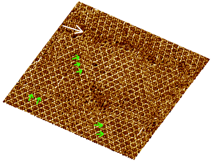
An unprocessed AFM image of a 2D crystal of a
membrane-binding protein, annexin V, is shown (680x680 nm).
Several stacking faults (green arrows) and a grain boundary
(white arrow) are visible.